New software from UHasselt and ZEISS shows in sharp detail how biomolecules move in cells.
Researchers from BIOMED UHasselt, in collaboration with the technology company ZEISS, have developed a software module that allows for even deeper and better examination of human cells using confocal microscopy. This software can create so-called heatmaps to visualize how biomolecules move within the cells. "This will significantly boost our understanding of cell biology and impact a wide range of biomedical and biological research," says Prof. Dr. Jelle Hendrix.
The software module from ZEISS and the team of Prof. Dr. Jelle Hendrix, based on patented research from UHasselt and LMU Munich, can be used for confocal microscopy.
In this type of microscopy, fluorescent dyes are used to color biomolecules, thus visualizing cells, the building blocks of the human body, in 3D. By mathematically analyzing these, researchers can deduce, for example, how many fluorescent biomolecules are present, whether they interact with their environment, and whether the biomolecules form functional networks. Such information is crucial for understanding how cells work and for determining what goes wrong in cells during certain diseases.
Great Added Value
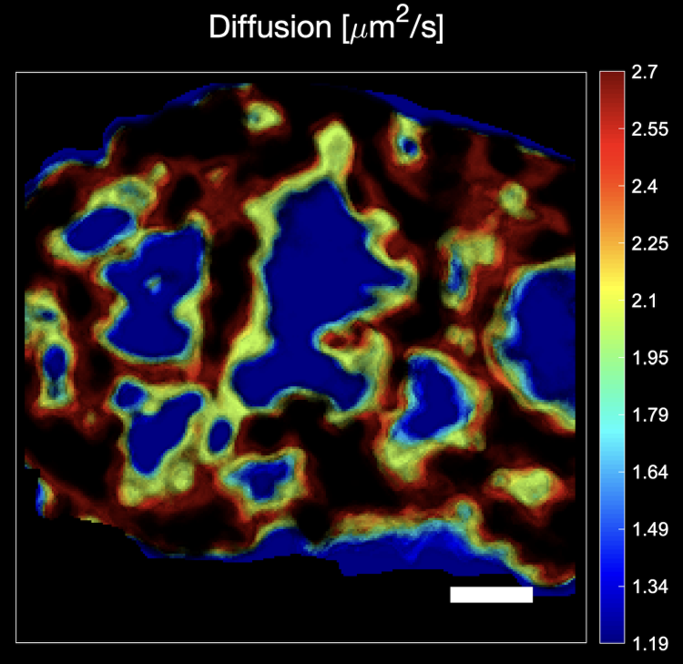
"Over the years, we have perfected these analyses, but we noticed that only niche researchers were using them. The newly developed software module now makes this type of analysis broadly accessible due to its user-friendliness, automation of the most complex calculations, and immediate display of analysis results while you are at the microscope," says Prof. Dr. Jelle Hendrix. "While most confocal microscopists previously could only map the composition of the cells during their recording sessions, they can now use the new software module to look sharply into the cell and see exactly which mechanisms are at play. Heatmaps, for example, show in detail where and when certain biomolecules come into contact with each other. This is a great added value in cell biology because knowledge of biomolecular contacts plays a key role in, among other things, drug development."
Market Leader
"ZEISS has been a market leader in this type of microscopy analysis for years. The great added value of this software module is that the behavior of biomolecules is not only quantified but also visualized. Microscopy images undergo a metamorphosis, so to speak. In this regard, the module is truly unique," says Dr. Cicerone Tudor, Applications Specialist at ZEISS Benelux.
Certificate of Excellence
The microscopy lab of the Biomedical Research Institute BIOMED at UHasselt recently received the prestigious ZEISS Labs@Location certificate. This quality mark is awarded by the high-tech company ZEISS to laboratories conducting top-level research using various types of microscopy.
"There are only about thirty laboratories worldwide that have this quality label," says Prof. Dr. Jelle Hendrix. "This certificate is a great recognition, especially within the development of the Health Campus Limburg DC. It allows us to test the latest technologies in microscopy in our lab and, as we are now, develop new solutions and products together with ZEISS."
Niek van Remortel, Head of Sales & Services at ZEISS Benelux, adds, "The ZEISS Labs@Location label is a win-win because we can give potential customers demonstrations on top equipment at UHasselt. This leads to greater visibility for both parties."
A Beautiful Journey
The UHasselt and ZEISS software has been on sale for a few weeks and will be rolled out to microscopy laboratories worldwide.
"The process from idea to patent and the final development of the software was a very exciting collaboration between BIOMED researchers and business developers, the Tech Transfer Office of UHasselt, and the microscopy company ZEISS. A fine example of knowledge transfer from the university to the outside world, one of the university's missions," says Dr. An Voets, Industrial Research Fund Business Developer at BIOMED.
"It is very gratifying to see an idea we played with a few years ago finally take shape, especially now that we see it can add so much value to microscopy research," concludes Jelle Hendrix.
For more information: Prof. Dr. Jelle Hendrix: jelle.hendrix@uhasselt.be
Image caption: Confocal heatmap indicating how fast biomolecules move at different positions in the cell nucleus.